Skip to content

Services

Specialty diagnostics, performed in your clinic.

Every service below is delivered on site at the primary-care hospital by Dr. Sikorski, a board-certified internist, with a written report to the referring veterinarian the same day.

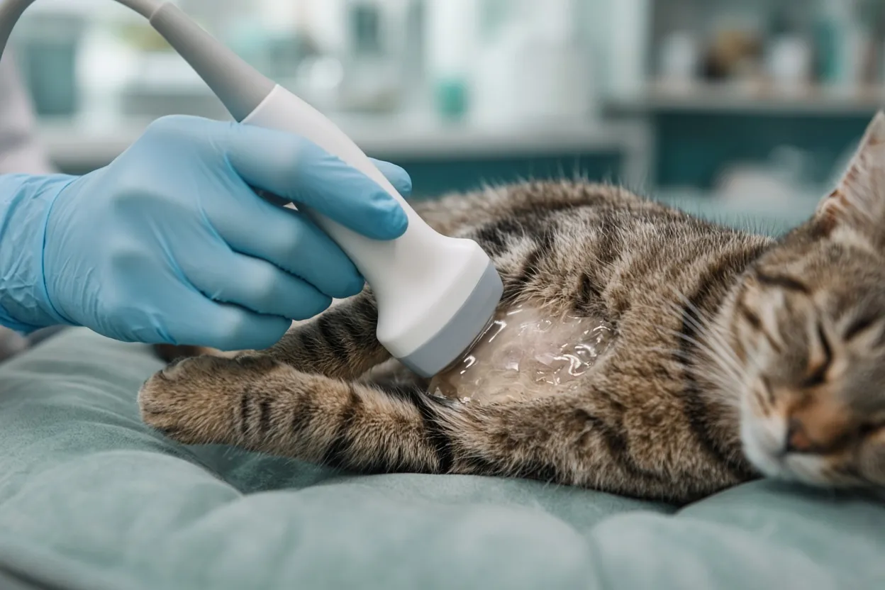
An ultrasound probe resting on the belly of a relaxed tabby cat

Abdominal ultrasound

A detailed, organ-by-organ look inside the abdomen when blood work and radiographs have not given the full answer.

Abdominal ultrasound examines each organ individually and in three dimensions: liver, gallbladder, spleen, kidneys, bladder, adrenal glands, pancreas, stomach and intestines, lymph nodes and the reproductive tract. The sonographer takes accurate measurements, records the internal appearance of each organ and watches real-time movement such as intestinal motility.

It is the usual next step for chronic vomiting or diarrhea, unexplained weight loss, abnormal liver or kidney values, suspected masses, urinary problems and many endocrine cases. When a sample is needed, ultrasound guides a fine needle aspirate or biopsy at the same visit.

Preparation: Withhold food for 12 hours (water is always fine). Juvenile patients and pets on insulin or food-dependent medication should not fast without a plan from the primary veterinarian.

Echocardiogram

An in-depth ultrasound of the heart to diagnose and stage heart disease and to make sense of a murmur.

An echocardiogram lets the sonographer see and measure each chamber of the heart, watch the valves, and track the direction and speed of blood flow with Doppler. It is the most accurate way to tell whether a pet has heart disease, how advanced it is, and which treatment makes sense.

Common reasons for referral include a newly detected murmur, coughing or breathing difficulty, exercise intolerance, an enlarged heart on radiographs, or breed screening. Conditions we evaluate include myxomatous valve disease, dilated cardiomyopathy, hypertrophic cardiomyopathy and hypertrophic obstructive cardiomyopathy. Prognosis is largely based on repeat echocardiography over time.

Preparation: No fasting is needed. A calm patient gives the best images, so discuss sedation in advance if your pet is anxious.

Small parts ultrasound

Focused scans of the neck, eyes, superficial masses and other small structures.

Small parts ultrasound is a targeted examination of structures outside the abdomen and chest: the thyroid and parathyroid glands, salivary glands, lymph nodes, the eye, superficial lumps and swellings. It answers a specific question quickly and can guide sampling of a mass without surgery.

Preparation: No special preparation. Clipping of the area is usually needed.

Musculoskeletal ultrasound

Real-time imaging of tendons, ligaments, muscles and joints.

Musculoskeletal ultrasound evaluates soft-tissue injuries that radiographs cannot show, such as tendon and ligament strains, muscle tears, joint effusion and swelling around a joint. Because it is a live image, the structure can be examined through its range of motion.

Preparation: No special preparation.

Ultrasound-guided fine needle aspirate

Cell samples from an organ or mass, collected under ultrasound guidance without surgery.

When ultrasound suggests inflammation, infection or cancer, a fine needle aspirate collects cells from the exact spot in question so a pathologist can define the process. It is performed at the same visit as the ultrasound, usually with only light sedation, and does not require a coagulation profile. Biopsies, which take a larger tissue core, do require a coagulation profile in advance.

If sampling is a possibility, we ask clinics to discuss consent with the owner before the appointment so the scan and the sample can happen together.

Preparation: Owner consent in advance. Coagulation profile for biopsies only.

Health check and pre-operative screening

A baseline abdominal or cardiac scan before anesthesia, for senior pets, or for peace of mind.

A screening ultrasound before surgery or as part of a senior wellness work-up can find problems early, when they are easiest to manage, and confirms that a patient is a good anesthetic candidate. It is also useful for breeds with known predispositions to heart disease or abdominal cancers.

Preparation: Same as abdominal ultrasound or echocardiogram, depending on the scan requested.

Internal medicine consultation

Board-certified case review, on site at your clinic or by telehealth.

Not every complex case needs imaging. Dr. Sikorski consults with primary veterinarians on difficult internal medicine cases: reviewing history and diagnostics, suggesting the most cost-effective next step, and building a treatment plan the clinic can carry out. Consultations can be scheduled alongside an ultrasound visit or as a standalone telehealth call.

Preparation: Send relevant history, lab work and imaging ahead of the appointment.

Conditions we help diagnose

From murmurs to mucoceles

Ultrasound and a specialist opinion are most valuable when blood work and radiographs have not given a clear answer. These are the problems we see most often.

Telehealth consultations

Wherever you are, we are. Dr. Sikorski is now scheduling telehealth consultations for veterinarians who want a specialist’s read on a case, a second opinion, or help planning the next diagnostic step.

Schedule a call

Ready to schedule?

Veterinary clinics can request an appointment online in about two minutes. Pet owners: ask your primary veterinarian to refer you to AIM.